
2026-10-15
Digital vs. conventional impressions: which is faster, more accurate and cheaper for a clinic
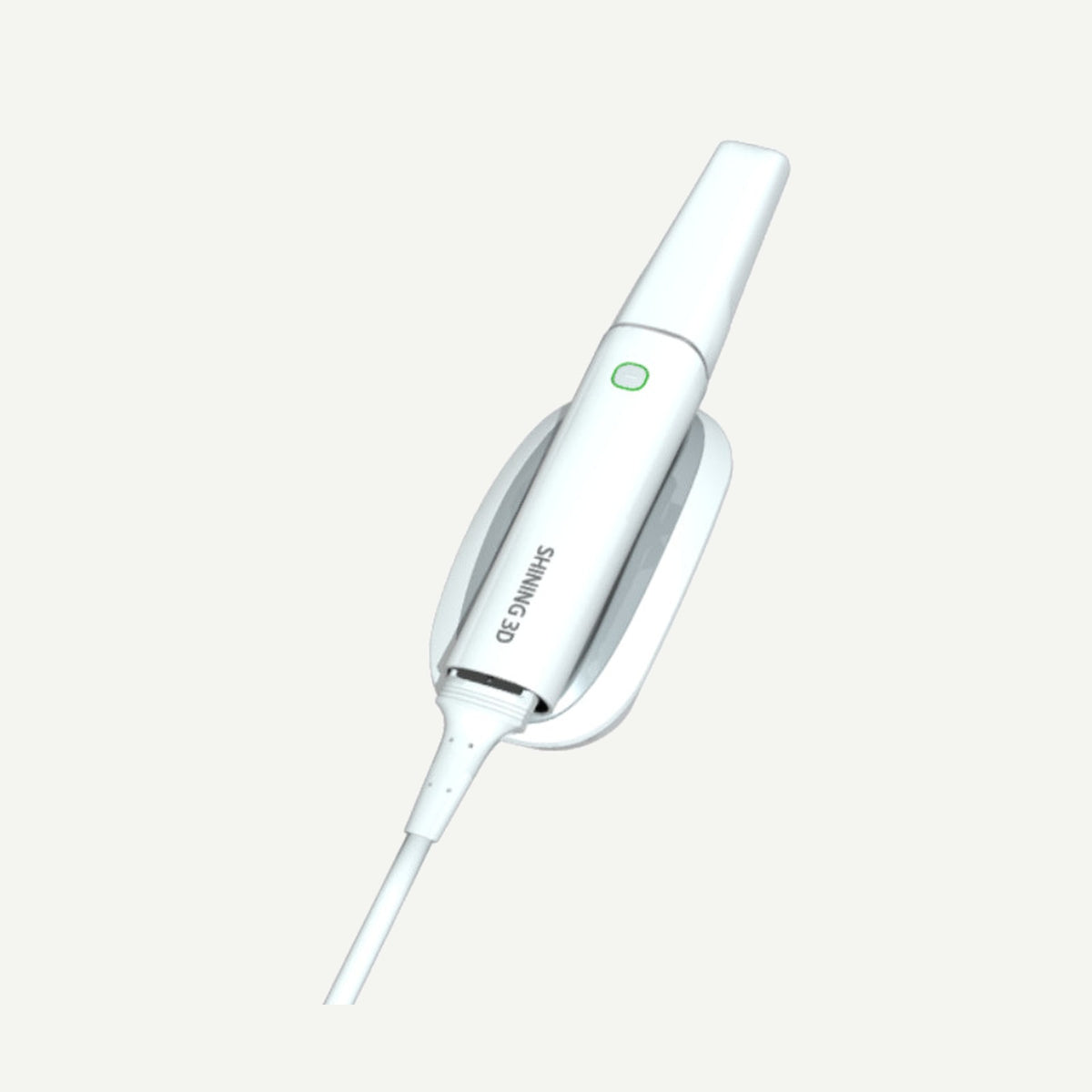
For most everyday work – single crowns, inlays, short bridges and orthodontics – a digital impression is faster, more comfortable for the patient and easier to send to the lab than a conventional silicone impression. The conventional impression still has its place: when the margin is deep subgingival, when there is bleeding, or when a full arch with several implants is needed without photogrammetry. Below we compare both methods in terms of time, accuracy, remakes, costs and communication with the lab – honestly, including the limitations.
Time: where you really save
The time a conventional impression takes is not just the setting time of the material in the mouth. You need to select a tray, mix the material, wait, remove it, evaluate it, repeat if necessary, disinfect and pack it. With a digital impression, most of the work happens right on the screen: you see the result immediately and can rescan only the missing area rather than the whole arch.
According to the manufacturer, the Aoralscan 3 scans one arch in about 25 s and both in about a minute. This is net scanning time – in a real appointment you add entering patient data, bite registration and checking the result. Still, according to the systematic review and meta-analysis by Manicone et al. (Journal of Prosthetic Dentistry, 2021), digital scanning for implant-supported restorations took less time than conventional impressions. A systematic review published in Frontiers in Oral Health in 2026 (Ramos-Morro et al.) also reports that digital impressions were consistently faster in the included studies, although the time measurement methodology varied between studies.
Accuracy: where the scanner is strong and where it is not yet
For short spans – single teeth, quadrants and short bridges – modern intraoral scanners achieve clinically sufficient accuracy. The 2026 review mentioned above, for example, cites a study in which the deviation of all tested scanners in quadrant impressions was below 100 µm.
For full arches the picture is more complex. In the same review, most of the included studies reported that conventional impressions were more accurate for full arches, especially over longer distances and in the posterior regions. That is why conventional scanning alone is often not enough for full-arch implant work (All-on-X) – the solution here is intraoral photogrammetry (IPG) with coded scan bodies, which the Aoralscan Elite offers. Read more in the article All-on-X scanning: when you need photogrammetry and coded scan bodies.
Limitations worth knowing in advance
- Subgingival margins. An optical scanner captures only what it "sees". If the preparation margin is deep subgingival and the sulcus is not retracted or is bleeding, a digital impression will not capture the margin. In such cases you need good gingival retraction and a dry field – or a conventional impression, where a lower-viscosity material can partly "push" under the gingiva.
- Learning curve. Scanning strategy (sequence, angles, managing the tongue and cheek) is learned, not innate. In the first weeks, appointments may take longer than usual until the team gets the hang of it. This is normal and worth including in the implementation plan.
- Mobile tissues and edentulous areas. In edentulous areas without clear landmarks it is harder for the scanner to "stitch" images, and a single scan does not reproduce the functional border of the mobile mucosa the way a functional impression with a custom tray does.
- Limited evidence on some metrics. There are few high-quality studies on remake rates and cost differences between the methods, so it is worth assessing these metrics using your own clinic's data.
Repeat impressions and remakes
With a conventional impression, some errors only become apparent in the lab: an air bubble at the margin, detached material, a distorted impression or a cracked stone model. The patient then has to be called back. When scanning, most of these problems are visible immediately – the missing area can be rescanned while the patient is still in the chair, and the preparation clearance and occlusion can be assessed on screen.
However, a digital impression does not fix a poor preparation or an unretracted margin – it just shows it sooner. The most reliable way to fewer remakes is to agree with the lab on what it must see in the scans (margin line, occlusion, opposing jaw, shade photos) and to check this systematically before sending.
Materials, logistics and other direct costs
Direct costs are easiest to compare by looking at what disappears and what is added.
| Cost item | Conventional impression | Digital impression |
|---|---|---|
| Materials | Impression material, mixing tips, trays, tray adhesive | No impression material needed; tips are reusable and sterilizable |
| Disinfection and packaging | Disinfection of each impression, packaging for shipment | Only the scanner and tips are disinfected |
| Logistics | Courier to the lab and back, stone models | The file is sent online; if needed, the lab prints the model |
| Equipment | No large initial investment | Scanner investment, computer, training, tip replacement |
| Time | Material setting, repeats, transport | Scanning and checking on screen, learning period |
What all this means for your clinic depends on the number of impressions per month and how often work has to be remade. We explain how to calculate this step by step in the article How much an intraoral scanner costs and how it pays for itself.
Patient comfort
For some patients, the conventional impression is the most unpleasant part of the appointment: the taste of the material, a full tray in the mouth and the gag reflex. In the meta-analysis by Manicone et al. (2021), patients rated digital scanning more favorably on all four assessed parameters – comfort, anxiety, nausea and perceived time. The 2026 Frontiers in Oral Health review, analyzing visual analogue scales, also found a clear patient preference for digital impressions. The authors note that study samples were small and participants were mostly young and fully dentate.
In practice, an added benefit is being able to show patients a 3D model of their teeth on screen. This makes it easier to explain the treatment plan, although the decision is, of course, driven by the clinical situation.
Communication with the lab
With a scanner, the lab receives the file within minutes of the appointment rather than the next day by courier. All Aoralscan models export open STL, OBJ and PLY files, so you can work with a lab that uses its own CAD software. Questions can be resolved while the patient is still in the chair: the technician looks at the scan and asks you to rescan the margin or add to the bite record.
Labs that still receive stone models or impressions digitize them with a lab scanner – so both paths merge into the same digital workflow. The most important thing is to agree in advance on the file format, the transfer channel and what the lab wants to see in each order.
When to still choose a conventional impression
- Deep subgingival margins when retraction and a dry field cannot be ensured.
- Full-arch work with several implants when the clinic does not have photogrammetry.
- Functional impressions of a completely edentulous jaw when the border of the mobile tissues needs to be reproduced.
- Temporarily – while the team is still learning and uses a proven method for more complex cases.
Many clinics work in a mixed way: they scan everyday cases and, in exceptional cases, use a high-quality addition silicone such as Vericom Vonflex S. You will find all impression materials in the Impression materials section.
Frequently asked questions
Is a digital impression more accurate than a silicone one?
For short spans – single teeth, quadrants, short bridges – studies show clinically sufficient and similar accuracy. For full arches, the conventional impression was more accurate in most studies, which is why photogrammetry is recommended for full-arch implant cases.
How long does it take to learn to scan?
A team usually masters the basic technique quickly, but fluency with full-arch scans and bite registration only comes with regular work. Plan slightly longer appointments for the first few weeks.
What if the margin is subgingival?
Ensure retraction and hemostasis so that the margin is visible and dry. If this is not possible, it is safer to take a conventional impression or combine the methods.
Will the lab accept my scans?
Aoralscan scanners export open STL, OBJ and PLY files that most CAD programs can open. Before you start, agree with the lab on the format and transfer channel.
Which scanner should I choose as a first step?
It depends on the indications: for general practice a model without photogrammetry is often enough, while implantology with full-arch work calls for a model with IPG. You will find a comparison in the articles Intraoral scanner – how to choose? and Aoralscan Elite, Elite Wireless or NEO.
Summary
For most everyday work, a digital impression saves time, reduces the risk of repeats and is more comfortable for the patient, while communication with the lab becomes faster. The conventional impression remains important for deep subgingival margins and functional impressions, and photogrammetry is best suited for full-arch implant cases. You will find SHINING 3D Aoralscan models in the Intraoral scanners section. Would you like to try scanning in your clinic? Contact IMMT or call +370 601 00030 – we will arrange a demo.